INTRODUCTION
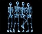
Radiography is the art and science of using radiation to provide images of the tissues, organs, bones, and vessels that comprise the human body. These images may be recorded on film or as a computerized image. Thus radiography is a paramedical professional course which is offered to individuals for performing diagnostic tests in medical treatment by the use of radiation.


Types of radiography
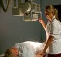
There are two types of radiography, diagnostic and therapeutic. Both need considerable knowledge of technology, anatomy and physiology and pathology to carry out their work. The ratio of diagnostic to therapeutic radiographers is ten to one. As a diagnostic radiographer one has to work mainly within the radiology and imaging departments of hospitals which includes a number of sections encompassing a wide range of different imaging modalities e.g. ultrasound, magnetic resonance imaging, nuclear medicine and, of course, X-rays.Diagnostic radiographers are able to undertake most investigations but may later specialise in one particular area. Diagnostic radiographers provide a service for most departments within the hospital including, accident and emergency, outpatients, operating theatres and wards. Most diagnostic radiographers carry out a range of procedures, they may specialise in techniques such as computerised tomography scanning, or magnetic resonance imaging which uses magnetic field and radio frequency waves to produce cross-sectional images of the body. Close liaison and collaboration with a wide range of other health care professionals is therefore vital.
Some of the imaging technology that a diagnostic radiographer may use is:
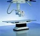
X-Ray - looks through tissues to examine bones, cavities and foreign objects.
Fluoroscopy - images the digestive system providing a real-time image.
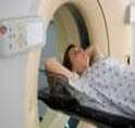
CT (Computed Tomography) - which provides cross-sectional views (slices) of the body.
MRI (Magnetic Resonance Imaging) - builds a 2-D or 3-D map of the different tissue types within the body.
Ultrasound - well known for its use in obstetrics and gynaecology. Also used to check circulation and examine the heart.
Angiography - used to investigate blood vessels
Therapeutic radiographers are increasingly known as radiotherapy radiographers who work closely with doctors, nurses, physicists and other members of the oncology team to treat patients with cancer. Radiotherapy radiographers may be involved in the care of the cancer patient from the initial referral clinic stage, where pre-treatment information is given, through the planning process, treatment and eventually post-treatment review (follow-up) stages. Thus they deliver doses of X-rays and other ionising radiation to patients, most of who are suffering from various forms of cancer.
Radiographers are in great demand in the modern health service, playing their part as key members of the healthcare team. By successfully completing undergraduate degree one will be able to practice as a qualified radiographer and put into practice your intellectual and caring capabilities. With the health services increasing by leaps and bounds the radiographers are constantly in demand in nursing homes, hospitals, diagnostic centres as well as super speciality hospitals. The more brilliant and experienced ones among can them get assignments in developed countries across the world. Radiographers have to work in diagnostic imaging department, intensive care unit, operating theatre, along with doctors and other hospital staff. The job of a radiographer also covers areas like medicine, research and teaching. Currently there are no direct entry routes into ultrasound. Most sonographers train as a radiographer then undertake an approved post-registration course. The field of nuclear medicine and photography are also open to x-ray technicians and radiographers. The following job profiles also comes under radiography
• Radio Diagnosis - It is the method of diagnosing the patient's illness with the help of X-ray, ultrasound or other such equipment.
• Radio Therapy - A therapy radiographer treat patients with tumour, using radiation in highly controlled conditions.
• Radiation Protection- Specialists in this area monitor the levels of radiation exposure and thereby ensure that no one is over-exposed to these radiations, this could prove to be extremely harmful.
Diagnostic radiography is a fast-moving and continually changing profession, and long-term career prospects include
• Management
• Research
• Clinical work
• Teaching
Although Wilhelm Röntgen is credited with the discovery of x-rays, he was almost certainly not the first to observe them, since they were readily produced using cathode ray devices. Many earlier scientists may have noticed but ignored such strange effects around their laboratories as glowing lights and foggy or overdeveloped photographic plates while experimenting with cathode rays, but probably dismissed or ignored them. It was Röntgen who recognized x-rays as a new type of radiation. Born in a small German village, Röntgen decided at an early age to study science, rather than follow his father as a cloth merchant. As a student, however, he preferred the outdoors to a classroom, and he was expelled from high school for assisting in a prank which had offended one of the instructors. Reputed to be insubordinate, Röntgen found the doors to the universities all but closed to him, and he was forced to apply to a local Technical School. Still, he completed his undergraduate studies in 1868, and in 1869 received his Ph.D. in philosophy.
Rontgen then moved to Zurich, Switzerland, and became an assistant to German physicist August Kundt (1839-1894), who introduced him to the world of physics.Röntgen received numerous accolades for his discovery, including the very first Nobel Prize for physics, but he invariably declined or donated any monetary prizes that would accompany his awards. He strongly believed that science belonged to everyone, and that all nations should benefit from its advances; he also refused to patent any facet of x-rays or their production. Thus, he was without substantial savings when the years following World War I brought hyperinflation to the German economy. He died in poverty in 1923 from intestinal cancer, probably caused by prolonged exposure to x-rays.
Horoscope - Career for Zodiac Signs
Thus if you have a mind of dedication for this noble profession then just check out these sun signs which are in favour of radiography career.
 Taurus
 Cancer
 Leo
Leo
 Virgo
Virgo
 Libra
Libra
 Scorpio
 Capricorn
 Aquarius
 Pisces
Eligibility : Click here for more information
Institutes : Some of the prominent institutions offering courses in Radiography can be had from the following links.. Click here for more information